


For more information about eye conditions, special tests and general eye health, visit the American Academy of Ophthalmology website.

| Name | What does it test? | Additional Information |
|---|---|---|
|
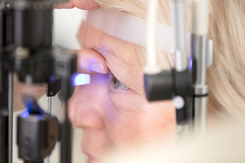
Tonometry |
Eye pressure |
Tonometry measures your eye pressure by determining how your cornea responds when an instrument (or sometimes a puff of air) presses on the surface of your eye. Eye drops are usually used to numb the surface of your eye for this test. |
|
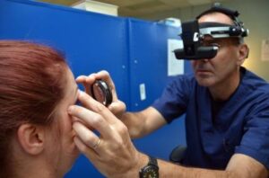
Gonioscopy |
Eye drainage angle |
Your eye’s drainage angle is the area where fluid drains out of your eye. During gonioscopy, you sit in a chair facing the microscope used to look inside your eye. You will place your chin on a chin rest and your forehead against a support bar while looking straight ahead.
The goniolens is placed lightly on the front of your eye, and a narrow beam of light is directed into your eye while your doctor looks through the slit lamp at the drainage angle. Drops will be used to numb the eye before the test. |
|
Ophthalmoscopy |
Optic nerve appearance |
With ophthalmoscopy, a high-powered lens is used to examine the optic nerve and retina. |
|
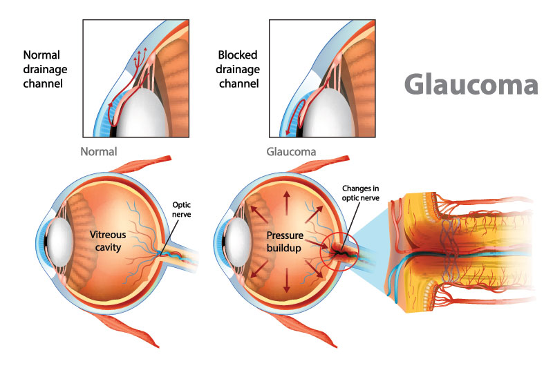
Visual field test |
Peripheral (side) vision |
A visual field test can help find certain patterns of vision loss and is a key way to check for glaucoma. It is very useful in finding early changes in vision caused by nerve damage from glaucoma.
This test is done seated at a bowl-shaped instrument called a perimeter. While you stare at the centre of the bowl, lights flash. Each time you see a flash, you press a button. A computer records the location of each flash and whether you pressed the button when the light flashed in that location. At the end of the test, a printout shows if there are areas of your field of vision where you did not see the flashes of light. This test shows if you have any areas of vision loss. Loss of peripheral vision is often an early sign of glaucoma. 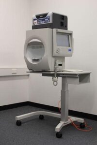 |
|
Optical coherence tomography (OCT) |
Thickness of the retina and optic nerve |
A healthy retina (the inner layer of tissue that lines the back of the eye) is only ¼ of a millimetre thick, but it contains multiple layers of specialized cells. One layer converts light into nerve signals, another processes the nerve impulses, while another transmits these processed impulses to the brain where they are interpreted. |